This comprehensive guide provides a hands-on approach to exploring human anatomy and physiology through cat dissection. Designed for students, it bridges theory with practical lab experiences, emphasizing structure, function, and dissection techniques. The manual is structured to enhance understanding of anatomical systems, making learning interactive and engaging for future healthcare professionals.
Overview of the Laboratory Manual
The Human Anatomy & Physiology Laboratory Manual, Cat Version is a comprehensive guide designed for students to explore anatomical structures through dissection. It features detailed diagrams, step-by-step procedures, and activities to enhance understanding of feline anatomy, which closely resembles human anatomy. The manual is organized into sections covering skeletal, muscular, nervous, circulatory, respiratory, digestive, urinary, and reproductive systems. Each chapter includes objectives, materials lists, and exercises to promote hands-on learning and critical thinking, making it an essential tool for anatomy and physiology education.
Importance of Cat Dissection in Anatomy and Physiology Studies
Importance of Cat Dissection in Anatomy and Physiology Studies
Dissection of cats in anatomy and physiology studies provides a practical understanding of mammalian anatomy, closely mirroring human structures. This hands-on approach allows students to identify and explore organs, bones, and tissues, enhancing their grasp of physiological processes. The similarity between feline and human anatomical systems makes cat dissection an effective tool for developing critical thinking and observational skills, preparing students for advanced studies in healthcare and biomedical fields. Additionally, it bridges the gap between theoretical knowledge and real-world application, fostering a deeper appreciation for human anatomy.
Structure and Organization of the Manual
The manual is organized into clear, logical chapters, each focusing on a specific anatomical system, such as skeletal, muscular, and nervous systems. Each chapter includes detailed descriptions, labeled diagrams, and step-by-step dissection guides, ensuring a comprehensive understanding. Practical exercises and lab activities are integrated to reinforce theoretical concepts, while the use of cat anatomy provides a relatable model for human physiology studies. The manual’s structured approach caters to students, offering a seamless transition from observation to hands-on exploration, enhancing learning efficiency and retention.

The Skeletal System
The skeletal system provides structural support and protection, comprising bones, cartilage, and ligaments. In cats, it mirrors human anatomy, making it ideal for comparative studies and dissection exercises.
Overview of the Skeletal System in Cats
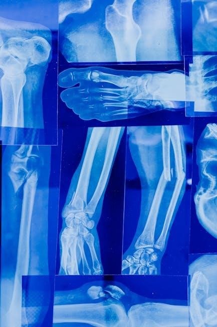
The feline skeletal system consists of 320 bones, providing structural support, protection, and facilitating movement. It includes the axial skeleton (skull, spine, ribs, sternum) and appendicular skeleton (limbs, shoulders, pelvis). Bones vary in shape and function, from the flexible vertebrae to the sturdy femur. The skeletal system also houses bone marrow for blood cell production and serves as an attachment site for muscles. Studying cat anatomy offers insights into human skeletal structure due to similarities, making it a valuable tool in laboratory settings. This system’s complexity allows for detailed examination of bone composition and joint mechanics.
Key Bones and Their Functions
In cats, key bones include the skull, vertebral column, rib cage, and limb bones. The skull protects the brain, while the vertebral column provides spinal support and flexibility. The rib cage shields internal organs like the heart and lungs. Limb bones, such as the scapula, humerus, radius, and ulna in the forelimbs, and pelvis, femur, patella, tibia, and fibula in the hindlimbs, enable movement and body support. The femur, the longest bone, is crucial for locomotion. These bones collectively facilitate movement, protection, and structural integrity in felines.
Lab Exercises for Identifying Bones
Lab exercises involve hands-on identification of feline bones, comparing them to human anatomy. Students examine skeletons, using diagrams and charts to label bones like the femur, humerus, and vertebrae. Activities include matching bones to descriptions, creating 3D models, and dissecting specimens to locate bones in context. Magnification tools help observe structural details, while quizzes assess identification skills. These exercises enhance understanding of bone functions and their roles in movement and support, bridging feline and human anatomical comparisons effectively.

The Muscular System
The muscular system in cats includes skeletal, smooth, and cardiac muscles, enabling movement, digestion, and circulation. Studying feline muscles provides insights into human anatomy and physiology.
Major Muscle Groups in Cats
Cats have distinct muscle groups, including flexors, extensors, epaxials, hypaxials, and limb muscles. Flexors enable limb bending, while extensors straighten them. Epaxial muscles run along the spine, aiding posture and movement. Hypaxial muscles support abdominal structures and respiration. Limb muscles, such as biceps and triceps, facilitate locomotion. These groups work synergistically, enabling cats to jump, climb, and maintain balance. Studying these muscles in cats provides valuable insights into human anatomy and physiology, as many muscle functions and structures are analogous between species.
Muscle Attachments and Movements
Muscle attachments in cats occur at specific origins and insertions, determining their range of motion. Origins are fixed points, while insertions are movable, enabling flexion, extension, and rotation. Tendons and ligaments connect muscles to bones, facilitating precise movements. In cats, muscles like the biceps femoris and gastrocnemius are specialized for agility and power. These attachments allow cats to perform unique movements, such as righting reflexes and climbing. Understanding these mechanisms helps students appreciate the functional anatomy of muscles in both feline and human systems, as many principles are conserved across species.
Practical Dissection of Muscles
Practical dissection of muscles involves carefully exposing and identifying muscle groups in cats. Proper preparation, including skin removal and tissue preservation, is essential. Students use scalpels and forceps to isolate muscles, tracing origins and insertions. This hands-on approach enhances understanding of muscle anatomy and function. Detailed illustrations and guides aid identification. Safety protocols, such as gloves and precise cuts, are emphasized to ensure effective learning and minimize tissue damage during dissection exercises.

The Nervous System
The nervous system in cats comprises the brain, spinal cord, and nerves, controlling voluntary and involuntary functions. It mirrors human anatomy, aiding students in comparative studies.
Structure of the Cat Brain and Spinal Cord
The cat brain consists of the cerebrum, cerebellum, and brainstem, regulating sensory, motor, and cognitive functions. The cerebrum processes sensory input, while the cerebellum coordinates movement. The brainstem connects the brain to the spinal cord, controlling involuntary actions like breathing. The spinal cord, part of the central nervous system, transmits nerve impulses between the brain and peripheral nerves. Its structure includes gray matter for processing and white matter for signal transmission, essential for reflexes and motor responses in cats.
Peripheral Nervous System in Cats
The peripheral nervous system (PNS) in cats includes nerves outside the brain and spinal cord, connecting the central nervous system to limbs and organs. It comprises the somatic nervous system, controlling voluntary movements, and the autonomic nervous system, regulating involuntary functions like heart rate and digestion. Nerves transmit signals, enabling reflexes, sensory perception, and motor responses. This system is vital for coordinating bodily functions and responding to environmental stimuli, making it a key focus in feline anatomy studies for understanding nervous system organization and function.
Nervous System Dissection Techniques
Nervous system dissection in cats requires precision to preserve delicate structures. Begin by carefully removing connective tissue to expose nerves and ganglia. Use scalpels and forceps to dissect nerve pathways, working under magnification if needed. Avoid cutting too aggressively, as nerves can tear easily. Gently separate nerves from surrounding tissues to trace their connections. Document findings with detailed sketches and notes. Proper technique ensures intact structures for study, aiding in understanding neural anatomy and its functional significance in both feline and human systems.

The Circulatory System
The circulatory system in cats transports blood, oxygen, and nutrients throughout the body, maintaining life and overall health through efficient vascular networks and cardiac function.
Heart Anatomy and Blood Vessels in Cats
The cat’s heart is a muscular, four-chambered organ with two atria and two ventricles. It pumps blood through arteries, veins, and capillaries. Arteries carry oxygen-rich blood away from the heart, while veins return oxygen-depleted blood. Capillaries facilitate gas and nutrient exchange. The heart’s structure includes valves ensuring one-way blood flow. During dissection, students can examine the coronary vessels supplying the heart muscle itself. Understanding feline cardiovascular anatomy aids in comparing it to human systems, enhancing physiological insights.
blood Flow and Circulation
Blood Flow and Circulation
Blood flow in cats is a continuous process essential for delivering oxygen and nutrients to tissues and removing waste products. The circulatory system ensures blood moves efficiently through arteries, veins, and capillaries. Arteries carry oxygen-rich blood away from the heart, while veins return oxygen-depleted blood. Capillaries enable exchange of gases and nutrients at the cellular level. The lymphatic system aids in returning fluids to the bloodstream. Studying feline circulation provides insights into human vascular physiology, highlighting similarities in blood flow mechanisms and overall systemic function.
Lab Activities for Examining the Circulatory System
In the lab, students engage in hands-on activities to examine the cat’s circulatory system, which mirrors human anatomy. Exercises include dissecting the heart to observe chambers and valves, identifying types of blood vessels, and tracing blood flow through the body. Students also measure blood pressure, analyze histological slides of blood vessels, and study capillary structures under a microscope. These activities enhance understanding of vascular physiology and prepare students for more complex studies in human health.
The Respiratory System
The cat’s respiratory system is studied to understand air movement and gas exchange, offering insights into human respiratory mechanics and related physiological processes effectively.
Lungs and Airway Structures in Cats
The cat’s respiratory system includes the trachea, bronchi, and lungs, which are essential for gas exchange. The trachea divides into primary bronchi, leading to the lungs. Feline lungs are divided into lobes, with the right lung having four lobes and the left having two. The lungs contain alveoli, tiny air sacs where oxygen diffuses into the blood. The diaphragm and intercostal muscles facilitate breathing. Studying these structures in cats provides valuable insights into mammalian respiratory anatomy, aiding in understanding human respiratory systems and related physiological processes effectively.
Mechanism of Breathing
Mechanism of Breathing
Breathing in cats involves the contraction and relaxation of the diaphragm and intercostal muscles; During inhalation, the diaphragm flattens, increasing chest cavity volume and drawing air into the lungs. Exhalation occurs as the diaphragm relaxes and the chest cavity shrinks. This process is essential for gas exchange, allowing oxygen to enter the bloodstream and carbon dioxide to be expelled. The mechanism is studied in detail during cat dissection, providing insights into respiratory physiology and its relevance to human anatomy and function.
Dissection of Respiratory Organs
Dissection of respiratory organs in cats involves carefully exposing the trachea, bronchi, and lungs. Students learn to identify structures such as the larynx, epiglottis, and diaphragm. The process requires precise incisions to avoid damaging delicate tissues. Observing the texture and color of lungs helps understand their function. This hands-on exercise provides a detailed understanding of respiratory anatomy, enhancing the ability to correlate feline structures with human respiratory systems for comparative learning.
The Digestive System
The digestive system in cats processes food into nutrients for absorption. It includes mouth, esophagus, stomach, and intestines, using enzymes and acids. Cats’ digestive systems resemble humans’, making them ideal for studying human digestive anatomy and physiology through dissection.
Organs of the Cat Digestive System
The cat digestive system consists of the mouth, esophagus, stomach, small intestine, and large intestine. The mouth begins digestion with teeth and saliva. The esophagus transports food to the stomach, which uses acids and enzymes to break it down. The small intestine absorbs nutrients, while the large intestine manages water and waste. Additionally, the liver, pancreas, and gallbladder support digestion by producing bile and enzymes. These organs function similarly to those in humans, making cats an excellent model for studying digestive anatomy.
Process of Digestion
Digestion in cats begins with ingestion, where food enters the mouth. Teeth chew food, and saliva moistens it. The esophagus propels the bolus to the stomach, where gastric juices break it down. The mixture enters the small intestine, where enzymes from the pancreas and bile from the liver and gallbladder further digest nutrients. Absorption occurs here, while the large intestine absorbs water and forms waste. This mirrors human digestion, making cat dissection a valuable learning tool for understanding the process in both species.
Lab Exercises for Exploring the Digestive Tract
Lab exercises involve dissecting a cat’s digestive system to identify organs such as the esophagus, stomach, small intestine, and large intestine. Students examine the pancreas and liver, observing their roles in digestion. Activities include tracing the flow of digestive enzymes and bile, and identifying structural features like gastric folds and intestinal villi. These hands-on exercises provide a detailed understanding of how the digestive system functions in cats, offering insights applicable to human anatomy and physiology.
The Urinary System
The urinary system in cats includes kidneys, ureters, bladder, and urethra. It filters waste, regulates electrolytes, and maintains fluid balance, mirroring human urinary functions, aiding comparative study.
Kidneys and Urinary Organs in Cats
Cats have two kidneys located near the lower back, filtering blood to produce urine. The renal cortex processes waste, while the medulla concentrates urine. Ureters transport urine to the bladder for storage. The bladder, a muscular sac, stores urine until release via the urethra. These organs mirror human urinary anatomy, aiding in comparative study. Their functions include waste removal, electrolyte regulation, and fluid balance, essential for overall feline health and physiological stability.
Urination Process
The urination process in cats involves the expulsion of urine from the bladder through the urethra. It is regulated by the nervous system, with the autonomic nervous system controlling involuntary bladder functions and the somatic nervous system enabling voluntary control. Stretch receptors in the bladder wall detect filling and signal the need to urinate. This process is crucial for eliminating waste and maintaining electrolyte and fluid balance, mirroring human urinary mechanisms and providing insights into comparative anatomy and physiology through dissection studies.
Lab Activities for Studying the Urinary System
Lab activities include dissection of the cat’s urinary system to identify and examine the kidneys, ureters, bladder, and urethra. Students observe the structure of nephrons under a microscope and trace the path of urine formation. Comparative analysis with human anatomy highlights similarities, such as the role of the kidneys in filtration. Hands-on exercises reinforce understanding of the system’s functions and prepare students for clinical applications in human physiology.

The Reproductive System
The reproductive system section examines cat anatomy through dissection, focusing on male and female structures. It highlights similarities with human systems, aiding in comparative learning for human anatomy students;
Male and Female Reproductive Organs in Cats
The male reproductive system includes the testes, epididymis, vas deferens, seminal vesicles, prostate, and penis, responsible for sperm production and delivery. The female system comprises ovaries, oviducts, uterus, cervix, vagina, and mammary glands, essential for ovulation, fertilization, and nurturing offspring. This section provides detailed dissection instructions, emphasizing anatomical structures and their functions. Understanding these organs aids in comparing feline and human reproductive systems, enhancing learning through hands-on exploration and visualization of physiological processes.
Reproductive Processes
In cats, reproduction involves complex physiological processes, including mating, fertilization, and embryonic development. Male cats produce sperm through spermatogenesis, while females undergo oogenesis to release ova. Hormonal regulation by estrogen and testosterone drives these processes. After mating, fertilization occurs in the oviduct, followed by implantation in the uterus. Gestation in cats lasts approximately 66 days, leading to birth. Lactation follows, nourishing kittens. This section details these processes, highlighting their importance in feline biology and their relevance to understanding human reproductive physiology through comparative anatomy.
Lab Exercises for Examining Reproductive Structures
Lab exercises involve detailed dissection of cat reproductive organs to identify key structures. Students examine the male reproductive system, focusing on the testes, epididymis, and vas deferens. For females, the ovaries, uterus, and vagina are studied. Dissections emphasize identifying anatomical features and understanding their functions. Proper use of dissecting tools and techniques is demonstrated to ensure accurate observation. These exercises provide hands-on experience, enhancing understanding of reproductive anatomy and its physiological processes, which are crucial for comparative studies in human anatomy and physiology;
This manual provides a comprehensive guide to understanding human anatomy through cat dissection. It emphasizes comparative anatomy, fostering deeper insights into structure-function relationships and practical dissection techniques.
This manual integrates anatomy and physiology through cat dissection, offering practical insights into skeletal, muscular, nervous, circulatory, respiratory, digestive, urinary, and reproductive systems. It provides detailed lab exercises, emphasizing structure-function relationships and dissection techniques. By comparing feline anatomy to human systems, students gain a foundational understanding of mammalian physiology. The manual bridges theory and practice, fostering critical thinking and hands-on learning for aspiring healthcare professionals and biologists.
Importance of Cat Dissection in Learning Human Anatomy
Dissection of cats is a valuable tool for understanding human anatomy due to their evolutionary similarities. Cats share many anatomical and physiological features with humans, making them an ideal model for studying mammalian systems. This hands-on approach enhances comprehension of complex structures and their functions. It also fosters critical thinking and practical skills essential for healthcare professionals. By comparing feline anatomy to human systems, students gain a deeper appreciation of shared biological principles and their clinical relevance.
